Search
15 results found with an empty search
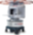
- Rurally Optimized MRI: Ultra-Low Field, Portability
Difficulty Conventional 1.5–3 T MRI assumes: Stable grid power (tens of kW) A shielded room and heavy infrastructure Cryogens and specialized service engineers Highly trained technologists and radiologists on site In many rural settings you instead have: Unreliable power, sometimes only generators or solar Limited space (small clinics, mobile vans, or health posts) Very few specialists and long referral times Patients who may travel hours–days for imaging Low-field (<1 T) and ultra-low-field MRI (ULF, typically ≤0.1 T) have been proposed specifically to loosen those constraints: permanent magnets, plug-in power, open geometries, simpler siting, and lower cost (Wald, 2019; Marques et al., 2019; Arnold et al., 2022). The question is: How far can we push low/ultra-low field and portability toward a truly rural-first design, not just a “smaller hospital” scanner? What ultra-low-field MRI already proves is possible Magnet field strengths and system simplification Key recent ULF milestones: 0.055 T brain scanner (permanent magnet, shielding-free) Liu et al. built a double-pole permanent magnet system (0.055 T) designed explicitly to be low-cost, low-power, and to operate without a full RF shielded room, using digital noise suppression instead (Liu et al., 2021). 0.05 T whole-body system Zhao et al. reported a 0.05 T whole-body scanner with linear gradients and demonstrated multiple clinical imaging protocols at that field, showing that even very low B₀ can still support diagnostically meaningful contrast when sequences and reconstruction are tuned properly (Zhao et al., 2024). 0.05 T MR angiography Ultra-low-field TOF-MRA at 0.05 T has been shown feasible, albeit with longer scan times and lower spatial resolution (Ultra-low-field MRA, 2024). A 2024 scoping review of ULF MRI emphasizes exactly these advantages: low power, smaller footprint, cheaper magnets, and the potential for portability and point-of-care use, while acknowledging trade-offs in SNR, spatial resolution, and susceptibility to environmental noise (Khan et al., 2024). Cost, power, and safety Low-field/ULF systems: Can use permanent magnets instead of superconducting ones, eliminating cryogens and greatly reducing maintenance (Wald, 2019; Anoardo et al., 2023). Have lower SAR, lower acoustic noise , and less stringent siting safety zones (Arnold et al., 2022). Can run on regular wall power or modest generators—critical for rural clinics (Wald, 2019; Murali et al., 2024). This is the physical foundation that makes “MRI in a village clinic” at least technically plausible. What portable MRI has already done in the real world Bedside and out-of-suite imaging A 0.064 T portable brain MRI system has been used: At the bedside in ICUs for critically ill patients who cannot be transported to the MRI suite (Yuen et al., 2022). With sensitivity around 94% for detecting brain lesions confirmed by 3 T MRI, although very small lesions are more easily missed at 64 mT (Arnold et al., 2022). Guallart-Naval et al. pushed this further, demonstrating a low-field extremity scanner that can operate indoors, outdoors, and in homes with a small footprint and modest shielding, effectively decoupling MRI from the hospital building entirely (Guallart-Naval et al., 2022). Relevance for rural settings Murali et al. argue that for low- and middle-income countries, low-field / portable MRI is one of the few viable paths to increasing scanner density, given constraints in capital, power, and MR-trained workforce (Murali et al., 2024). Taken together, existing portable systems show that: Wall-plug or generator-powered MRI is feasible. A scanner can be wheeled to the patient, or carried in a van to a community. You don’t need a full-size shielded suite if you manage noise cleverly. But most current devices are still priced and serviced with high-income hospitals in mind, not village clinics. Design principles for a rural-first ULF portable MRI Think of this less as “a shrunk 3 T scanner” and more as a diagnostic appliance for a very specific set of questions (stroke vs no stroke? mass vs no mass? spinal compression?), tuned to rural constraints. Magnet and field strength Conceptual choice: ~0.05–0.1 T permanent magnet. 0.05–0.06 T has been demonstrated for brain and whole-body imaging with permanent magnets (Liu et al., 2021; Zhao et al., 2024). A C-shaped or double-pole magnet with an open front allows seated or partially reclined positioning and easier patient access. A Halbach or multi-ring permanent array can concentrate field in the imaging volume and reduce stray field (Anoardo et al., 2023). Design goals: No cryogens Total magnet + gradient assembly mass < ~500–700 kg (so it can be van-mounted or rolled over modest surfaces) Homogeneous field over a head-sized or extremity-sized volume Gradients and RF Low-power, water- or air-cooled gradients optimized for head and spine, not full body. Use insights from recent work on dual-polarity gradient schemes that push SNR and speed at ULF by smarter sequence design rather than brute force hardware (Lau et al., 2023). A limited number of RF coils : One head coil (receive array if budget allows) One extremity coil (knee / ankle / wrist) Integrated small RF shield around the magnet only , avoiding the need for a dedicated room (Liu et al., 2021). Power, cooling, and siting Operate from standard 110/220 V, <2–3 kW draw, compatible with clinic power or a small generator (Wald, 2019). Air-cooled electronics, no water chiller. No fixed room build-out : the system sits on a wheeled base or in a van; RF shielding is either localized (around magnet) or achieved with active noise cancellation. Rural-specific tweak: overspec the system for voltage fluctuations , with a battery buffer or UPS integrated into the base. Software, reconstruction, and AI At ULF, the biggest bottleneck is SNR and resolution, not hardware cost. That’s where reconstruction and AI matter. Fast, ULF-tuned sequences (e.g., T2-weighted, FLAIR-like, diffusion-simplified) focused on yes/no clinical questions (Arnold et al., 2022; Khan et al., 2024). Image-to-image deep learning for denoising and super-resolution: Islam et al. showed that a GAN-based model (LoHiResGAN) can map 64 mT images to synthetic 3 T-like images, substantially improving perceived quality (Islam et al., 2023). Edge device does basic reconstruction; heavier AI runs in the cloud or on a central server when connectivity exists. Built-in tele-radiology : one-click upload to a reading hub. For rural deployment, the UI should look more like a tablet app than a hospital console: exam presets (“Stroke screen”, “Brain mass screen”, “Pediatric hydrocephalus”), simple traffic-light quality indicators, and automatic anonymization for remote reads. Clinical protocol philosophy Instead of “all the sequences,” aim for 3–5 short protocols : Acute neuro protocol (10–15 min) Axial T2 / FLAIR-like Basic diffusion (if feasible at ULF)Purpose: large infarcts, hemorrhage, mass effect, hydrocephalus. Chronic neuro protocol (15–20 min) T1-weighted structural T2 / FLAIR-likePurpose: tumor follow-up, white-matter disease, moderate atrophy. Spine or extremity protocol (10–15 min) Sagittal + coronal T2Purpose: cord compression, fracture, osteomyelitis, joint effusions. Scan times and contrasts are guided by what has already been achieved at ~0.05–0.064 T in research and early clinical systems (Zhao et al., 2024; Yuen et al., 2022; Guallart-Naval et al., 2022). Human factors and training One-week training curriculum for rural clinicians or radiographers: positioning, safety, basic troubleshooting. Extensive on-screen guidance and remote support chat/video. Design the physical form so that: It fits through a standard clinic door. The patient can be imaged on a simple stretcher or wooden bed, not a hospital gurney. Local artisans can build ramps or platforms if the scanner is van-mounted. A preliminary conceptual design: “Village MRI Cart” Here’s one concrete concept you could sketch for a design brief or early prototype. Hardware snapshot Field strength: 0.06 T permanent double-pole magnet (head-optimized) Geometry: Open front “drum” that a patient’s head or knee can slide into Magnet + gradient + RF in a cylindrical module ~80 cm diameter Base: Motorized cart with four large wheels (for uneven clinic floors) Integrated 3–5 kWh battery pack and power electronics Mass target: ~500 kg total Cooling: Forced air, filters that can be cleaned locally Shielding: Local RF cage around magnet with modular panels, plus digital noise filtering Software and workflow Set-up Plug into wall or generator; system auto-checks line quality and falls back to battery if unstable. Tablet interface boots a guided workflow. Patient exam Operator selects preset (“Stroke screen”). UI shows where to place the head and how to center it with simple visual markers. Short scout scan checks positioning and noise; system gives “OK / re-position / too noisy” feedback. Reconstruction Raw k-space reconstructed locally. On-device denoising + compressed sensing. When online, images are uploaded for cloud AI enhancement (e.g., LoHiRes-style network) and remote radiologist reading (Islam et al., 2023). Reporting Rural clinician gets a simple structured report and key images back (e.g., within a few hours), plus triage recommendation (“urgent transfer”, “routine follow-up”, etc.). Deployment model for rural regions Hub-and-spoke : One scanner per district hospital, plus one van-mounted unit that travels on a weekly schedule to peripheral clinics. Maintenance: Local technician trained for basic issues; remote diagnostics and scheduled annual visits by manufacturer. Cost target: Capital cost well below standard 1.5 T (Wald, 2019; Murali et al., 2024 suggest an order-of-magnitude reduction is plausible with permanent magnets and simpler infrastructure). Where the research gaps still are Even with all this, several things are not solved yet: Evidence base at ULF is still thin for many pathologies compared with 1.5–3 T (Arnold et al., 2022; Khan et al., 2024). Robustness outside hospitals —think dust, humidity, RF noise from local industry—needs more real-world trials (Guallart-Naval et al., 2022). AI models trained mostly on high-income populations may not generalize perfectly to rural demographics and co-morbidities (Islam et al., 2023). But the combination of: Permanent-magnet ULF hardware , Portable form factors , and Modern reconstruction / AI is already far enough along that “MRI in a village clinic” is no longer science fiction—it’s an engineering, regulatory, and business-model problem. References Arnold, T. C., et al. (2022). Low-field MRI: Clinical promise and challenges. Journal of Magnetic Resonance Imaging . Arnold, T. C., et al. (2022). Sensitivity of portable low-field magnetic resonance imaging for detecting brain lesions. Scientific Reports . Guallart-Naval, T., et al. (2022). Portable magnetic resonance imaging of patients indoors, outdoors and at home. Scientific Reports . Islam, K. T., et al. (2023). Improving portable low-field MRI image quality through image-to-image translation using paired low- and high-field images. Scientific Reports . Khan, M., et al. (2024). Applications, limitations and advancements of ultra-low-field magnetic resonance imaging: A scoping review. Surgical Neurology International . Lau, V., et al. (2023). Pushing the limits of low-cost ultra-low-field MRI by dual-polarity gradient encoding. Magnetic Resonance in Medicine . Liu, Y., et al. (2021). A low-cost and shielding-free ultra-low-field brain MRI scanner. Nature Communications, 12 , 7238. Marques, J. P., & Simonis, F. F. J. (2019). Low-field MRI: An MR physics perspective. Journal of Magnetic Resonance Imaging . Murali, S., et al. (2024). Bringing MRI to low- and middle-income countries. NMR in Biomedicine . Ultra-low-field magnetic resonance angiography at 0.05 T. (2024). NMR in Biomedicine . Wald, L. L. (2019). Low-cost and portable MRI. Journal of Magnetic Resonance Imaging . Yuen, M. M., et al. (2022). Portable, low-field magnetic resonance imaging enables bedside assessment of critically ill patients. Science Advances . Zhao, Y., et al. (2024). Whole-body magnetic resonance imaging at 0.05 Tesla. Science .
- Policies that Hinder the Development of Neurotechnology in Rural Areas
When people talk about “neurotechnology,” they usually picture gleaming MRI scanners, EEG caps, neuromodulation devices, or AI tools reading brain scans in big-city hospitals. But for many rural communities, the real story is less about hardware and more about policy: who gets paid, who is allowed to practice, what infrastructure gets funded, and who sits at the procurement table. Those rules can quietly decide whether neurotechnology ever makes it to a village clinic or a critical-access hospital. 1. Financing rules that make neurotech a losing bet Most advanced neurotechnology is expensive to buy, run, and maintain. Rural hospitals and clinics typically operate on very thin margins, so policy-driven payment rules can make or break any investment. In the U.S., federal telehealth and reimbursement policy shows how this plays out. A 2024 National Rural Health Association policy brief notes that Medicare historically limited telehealth coverage based on geography and “originating site” rules, and only reimburses a finite list of services that have an in-person equivalent (identified by specific billing codes) (National Rural Health Association, 2024).That’s fine if you’re billing for a standard neurology consult—but much less clear for rural teleradiology, remote EEG interpretation, or continuous neuro-monitoring. The same brief emphasizes that rural health clinics and federally qualified health centers must still pay for brick-and-mortar overhead and digital infrastructure, while reimbursement often doesn’t fully account for those extra costs (National Rural Health Association, 2024). In parallel, the Rural Health Information Hub points out that ambiguous Medicaid rules and inconsistent private-payer coverage have made reimbursement one of the historic barriers to rural telehealth (Rural Health Information Hub, 2023).When the financial math doesn’t work, rural administrators understandably hesitate to bring in a new EEG system, low-field MRI, or tele-neurology platform—no matter how “innovative” it is. In low- and middle-income countries (LMICs), the dynamic is similar but starker. Lancet-linked work on imaging in LMICs highlights chronic under-investment, high equipment costs, and lack of long-term maintenance budgets as core barriers to MRI and CT deployment (Frija et al., 2021). Without sustained capital and operating funding written into national plans, neuroimaging remains concentrated in big referral hospitals, leaving rural clinics to manage stroke, epilepsy, and dementia essentially “blind.” 2. Licensing, credentialing, and the tele-neurology maze Even when the technology exists, policy can make it hard for specialists to actually use it for rural patients. Both RHIhub and NRHA describe how telehealth providers are usually required to hold a license in every state where their patients live, with complex, time-consuming credentialing processes for each hospital (Rural Health Information Hub, 2023; National Rural Health Association, 2024). Interstate compacts help, but coverage is patchy, and rural hospitals often lack the administrative staff to navigate credentialing and privileging requirements for multiple neurologists or neuroradiologists (National Rural Health Association, 2024). For frontier communities that could be served by a tele-stroke neurologist or remote epilepsy specialist reading EEGs, licensing and credentialing rules effectively throttle access. There’s evidence that these barriers matter for neurological care specifically. Studies of rural multiple sclerosis and neurology care in North America report fewer neurologist visits, fewer disease-modifying prescriptions, and lower perceived care quality among rural patients, even when telehealth is available (Cochran, McGinley, & Palmer, 2024). Telehealth is cited as a promising fix—but its impact is limited by exactly these policy barriers: licensing complexity, digital literacy, and infrastructure gaps. 3. Infrastructure policies: broadband and power as gatekeepers Neurotechnology is data-hungry. MRI images, EEG traces, or remote neuromodulation logs are useless if they can’t be transmitted safely and reliably. RHIhub estimates that about 28% of people in U.S. rural areas lack access to high-speed broadband, with even worse numbers on Tribal lands (Rural Health Information Hub, 2023). The NRHA brief notes that roughly one-third of rural Americans lack sufficient broadband, and older adults—who are more likely to need neuro care—are especially underserved (National Rural Health Association, 2024).Without strong connectivity, rural hospitals can’t reliably upload MRI scans for tertiary review, stream real-time telestroke consults, or support cloud-based AI tools. Globally, infrastructure policies play a similar role. RSNA’s 2024 report on imaging in LMICs describes how radiology departments are embedded in an “ecosystem” that requires dedicated space, stable electricity, local maintenance capacity, and future-proofing for upgrades (Silverberg, 2024). In many rural regions, national grid investments, facility planning standards, and biomedical engineering training policies haven’t caught up, so even donated or procured equipment sits idle. 4. Equipment procurement and planning that bypass rural voices Even where money and electricity exist, how equipment is chosen and procured matters. The RSNA report notes that in many LMICs, national or provincial procurement processes purchase large imaging units from foreign vendors, often with long delays, expiring warranties, and little input from radiologists on what’s actually needed (Silverberg, 2024).Bureaucratic rules can lock rural hospitals into one-size-fits-all tenders that favor high-end machines for urban centers, instead of portable or lower-field systems more appropriate for district hospitals. At the policy level, this shows up as the absence of clear, rural-sensitive technology assessment frameworks. Lancet-linked work on imaging in LMICs flags three key policy failures: lack of national investment plans that prioritize primary-care-level imaging, high equipment and maintenance costs, and difficulty operating complex equipment safely in under-resourced settings (Frija et al., 2021). That combination pushes ministries to buy fewer, high-end machines for cities rather than many simpler systems distributed across rural regions—slowing the spread of neuroimaging and other neurotech. China’s experience with rural primary health care shows how even pro-equipment reforms can leave gaps. A 2024 study on remote western regions found that policy interventions did increase the supply of basic medical equipment in rural primary care facilities, but the distribution remained uneven and still lagged behind population needs (Shan et al., 2024). In other words, policy can grow the equipment “pie” while still leaving rural communities with the smallest slices. 5. Workforce and training policies: no specialists, no neurotech Neurotechnology doesn’t run itself; it needs neurologists, neuroradiologists, neurosurgeons, EEG technicians, and biomedical engineers. Workforce policy has been slow to respond. The American Academy of Neurology Workforce Task Force projected more than a decade ago that demand for neurologist services would outstrip supply in most U.S. states by 2025 (Freeman et al., 2013). More recent work shows just how unequal the distribution is: one study reported roughly 6.2 neurologists per 100,000 people in urban U.S. counties versus only 1.2 in rural counties (Amiri et al., 2024). For neurotechnology, that means even when an MRI or EEG machine is present, there may be no specialist to interpret results or supervise advanced procedures. Policy levers—graduate medical education caps, rural residency incentives, scope-of-practice rules, and task-shifting regulations—directly shape this landscape. Yet many systems still tightly restrict who can interpret neuroimaging or EEGs, and underutilize community health workers or non-physician clinicians in screening and follow-up roles. Studies of village clinicians in rural China, for example, show that clinicians completed only about 26% of recommended diagnostic questions and exams and correctly diagnosed around 20% of standardized heart-disease cases, despite being the main frontline providers (Guo et al., 2020). Without parallel investments in training, supervision, and task-sharing policy, deploying neurotechnology risks widening the “know-do” gap rather than closing it. 6. Regulatory and ethical frameworks struggling to keep up Finally, some policies hinder neurotechnology precisely because they weren’t designed with it in mind . Portable and ultra-low-field MRI, point-of-care EEG, and brain–computer interface (BCI) systems challenge older assumptions about where brain data is collected and who controls it. A 2021 NeuroImage paper on highly portable MRI in remote, low-resource settings argues that these systems raise under-examined ethical, legal, and social issues—around informed consent, data governance, incidental findings, and community engagement—that existing regulations don’t fully address (Shen et al., 2021).Where national ethics and device-approval frameworks haven’t yet adapted, local review boards may default to conservative decisions, slowing field-based neuroimaging projects in exactly the rural areas that could benefit most. Similarly, privacy and interoperability regulations intended to protect patients can become operational barriers if rural hospitals lack the technical staff and secure infrastructure to meet stringent requirements for transmitting large neuroimaging files or continuous neural data streams (National Rural Health Association, 2024; Rural Health Information Hub, 2023).The result is a paradox: neurotech is “approved” in theory but practically unusable in many rural facilities. Where policy could move next The literature doesn’t say that neurotechnology and rural equity are incompatible. It says the opposite: when rural-sensitive policies are in place—like broadband investment programs, telehealth reimbursement parity, rural training tracks, and procurement frameworks that prioritize primary-care-level imaging—technology can dramatically improve neurological outcomes and be cost-effective in the long run (Silverberg, 2024; Frija et al., 2021; Shan et al., 2024). But the current policy environment still often treats neurotechnology as an urban specialty luxury. Until financing, licensing, infrastructure, workforce, and regulatory frameworks are rewritten with rural contexts in mind, even the most “disruptive” devices will keep bouncing off the same invisible walls. For anyone working on rural-optimized neurotechnology—low-field MRI, portable EEG, GBCI, BCIs, or teleneurology—reading the policy literature is almost as important as reading the engineering papers. The barriers are legal and economic as much as they are technical, and changing them is part of the R&D process, not an afterthought. References Amiri, S., et al. (2024). Racial, ethnic, and rural disparities in distance to neurologists and primary care physicians among individuals with Alzheimer’s disease and related dementias. [Journal of neurology/health services research] . Cochran, J., McGinley, M., & Palmer, K. (2024). Unique health care delivery considerations in rural America. International Journal of MS Care . Freeman, W. D., Vatz, K. A., Griggs, R. C., & Pedley, T. (2013). The Workforce Task Force report: Clinical implications for neurology. Neurology, 81 (5), 479–486. Frija, G., et al. (2021). How to improve access to medical imaging in low- and middle-income countries? EClinicalMedicine, 41 , 101136. Guo, W., Sylvia, S., et al. (2020). The competence of village clinicians in the diagnosis and management of heart disease in rural China. The Lancet Regional Health – Western Pacific, 1 , 100004. National Rural Health Association. (2024). Impact of telehealth policy on rural health access (Policy brief). Rural Health Information Hub. (2023). Barriers to telehealth in rural areas. In Rural Telehealth Toolkit . Shan, L., Gan, Y., Yan, X., Wang, S., Yin, Y., & Wu, X. (2024). Uneven primary healthcare supply of rural doctors and medical equipment in remote China: Community impact and the moderating effect of policy intervention. International Journal for Equity in Health . Shen, F. X., et al. (2021). Emerging ethical issues raised by highly portable MRI research in remote and resource-limited international settings. NeuroImage, 225 , 117480. Silverberg, M. (2024). How radiologists overcome barriers to provide imaging in low to middle income countries. RSNA News .
- All Prototypes for NeuroGut Tracker Finished!!!
Check our main website for details.
- NeuroGut-Daily Prototype Finished!
NeuroGut-Clinical on its way
- Introducing 2 Versions of NeuroGut Tracker
We've designed two versions of the NeuroGut Tracker: NeuroGut-Clinical and NeuroGut-Daily While both ensures high wearability and lightness, NueroGut-Clinical maximizes accuracy, while NeuroGut-Daily maximizes portability. STAY TUNED!!!!
- GI Treatment in Rural Areas & Additional Dementia Caring Cost for Neurologically-linked GI Commorbidities
When the Gut and Brain Both Need Help—but the Clinic Is Far Away Imagine an 82-year-old farmer in a rural county who has Alzheimer’s and chronic constipation, wakes up some days with reflux, and occasionally chokes on food. None of those issues on their own sound dramatic. But together they can mean ER trips for GI bleeding, aspiration pneumonia, or dehydration—while a specialist who understands both gut and brain is more than an hour’s drive away. That’s the lived reality behind a growing but still under-discussed problem: GI treatment in rural areas and the extra dementia care costs driven by neurologically linked GI comorbidities. 1. GI care in rural areas: distance, delay, and workarounds At a systems level, the first barrier is brutally simple: there often is no gastroenterologist nearby. A 2025 analysis of U.S. physician workforce data found that almost 50 million Americans—disproportionately in rural counties—must travel at least 25 miles to see a gastroenterologist, and over two-thirds of U.S. counties have no gastroenterologist at all. About 80% of these “GI-desert” counties are non-metropolitan, older, poorer, and less insured than counties with a specialist. Where there are no GI doctors, rural systems improvise. A qualitative study of colonoscopy provision in rural Oregon showed that much of the endoscopy work is done by primary-care physicians and general surgeons who scope their own patients or accept referrals, often for Medicaid populations. Providers described multilevel barriers—travel, time off work, fear of costs, and shrinking numbers of primary-care endoscopists—as threats to sustaining access. Earlier work showed that rural residency is associated with fewer colonoscopy providers overall, longer waits, and decreased utilization, even when patients are eligible for screening. All of this matters because chronic GI disease is not “cheap.” In the U.S., chronic GI and liver diseases account for heavy hospitalization and cost burdens, including billions annually for upper GI bleeding and hundreds of thousands of admissions. For older adults, common issues like dyspepsia, GERD, constipation, fecal incontinence, and motility problems are highly prevalent and often more severe or more complicated than in younger patients. When those problems go underdiagnosed or undertreated in rural settings—because there’s no endoscopist, no motility lab, no easy follow-up—they quietly accumulate risk. 2. The gut–brain axis: why GI problems are not “separate” from dementia Over the last decade, the microbiota–gut–brain axis has shifted how we think about GI disease and neurodegeneration. Reviews in Signal Transduction and Targeted Therapy and related journals describe a bidirectional network in which gut microbes, immune signaling, and the vagus nerve influence microglial activation, neuroinflammation, and synaptic function, making the gut a real lever for neurodegenerative disease pathways. Epidemiologic data are now catching up. A large prospective cohort study in American Journal of Preventive Medicine found that eleven digestive system diseases—including cirrhosis, peptic ulcer disease, and inflammatory bowel disease—were significantly associated with higher incident dementia risk after adjusting for genetics and vascular factors. A meta-analysis focusing specifically on inflammatory bowel disease (IBD) reported that IBD is associated with increased all-cause dementia risk, though estimates vary and more prospective data are needed. Other work links slower, more “everyday” GI problems to brain outcomes: research presented at the Alzheimer’s Association International Conference in 2023 showed that less frequent bowel movements in mid- to late life were associated with faster cognitive decline. So when a rural dementia patient has chronic constipation, reflux, IBD, dyspepsia, or chronic GI bleeding, those aren’t just side quests. They sit right inside the gut–brain loop that influences inflammation, nutrition, mood, and ultimately dementia trajectory. 3. Dementia is already expensive—multimorbidity makes it worse Even without GI problems, dementia is one of the most expensive conditions in the world. Hurd and colleagues estimated that in 2010, dementia added roughly $33,000 per person per year in extra health-care costs in the United States, with total national costs (including informal care) in the $159–215 billion range. Global modeling suggests direct costs could reach around $2 trillion by 2030 if trends continue. Claims–based studies consistently show that people with Alzheimer’s disease (AD) or related dementias have more comorbid medical conditions and higher Medicare expenditures than similar older adults without dementia.When you add Medicaid, the spending gap widens further: in one ethnically diverse cohort, annual Medicaid expenditures for people with dementia averaged about $50,000 versus $22,000 for those without dementia, with combined Medicare–Medicaid spending nearly double. Recent work in England found that “high-cost” AD dementia subgroups—characterized by greater frailty and more cardiometabolic comorbidities—spent 1.3–1.7 times the average per-person cost, with social care dominating the bill.Behavioral symptoms, agitation, and other complications also significantly increase health-care utilization and cost. Taken together, the literature is clear on one point: comorbidities are cost multipliers in dementia. 4. GI comorbidities as hidden cost drivers in dementia care Where the evidence is thinner—but emerging—is the specific role of GI comorbidities in those costs. Dysphagia (swallowing difficulty) is a good example. In general geriatric populations, dysphagia is associated with higher hospital and municipal care costs; one Danish study estimated that older adults with dysphagia incurred roughly €3,700 more in hospital costs and over €6,000 more in municipal health-care costs per year than those without, even after adjusting for age and comorbidities. Systematic reviews in Alzheimer’s disease show that dysphagia prevalence rises dramatically with disease severity and is linked to aspiration pneumonia, malnutrition, institutionalization, and increased overall health-care costs. A 2023 ICU-based study of older adults with dementia found that those with dysphagia had significantly higher 90- and 180-day mortality, more pressure injuries, more aspiration pneumonia, and were more often discharged to nursing facilities rather than home—all of which are cost-intensive outcomes. Other GI problems create similar financial drag. Chronic GI and liver diseases generate high rates of hospitalization and billions in direct costs annually. In older adults with dementia, colonoscopy appears technically feasible but is associated with longer length of stay, more non-GI complications (like kidney injury and pneumonia), and higher hospital costs compared to patients without dementia. If you overlay that on the earlier dementia-cost picture, the story becomes: Dementia itself is expensive; Multimorbidity, frailty, and complications push selected subgroups into very high-cost tiers; GI comorbidities—especially dysphagia, IBD, bleeding, and motility disorders—are common in older adults, bi-directionally linked to brain health, and associated with greater hospital use, institutionalization, and supportive care needs. There are not yet many papers that compute “the exact extra dollars of dementia care attributable to neurologically linked GI comorbidities.” But the pieces we do have strongly imply that these conditions add a substantial incremental burden, particularly via hospitalizations, long-term care placements, and caregiver time. 5. Why rural GI gaps amplify dementia costs Now put all of this back into a rural setting: Fewer specialists and longer travel distances mean delayed diagnosis of dysphagia, IBD, or GI bleeding. That can mean presenting late with severe anemia, perforation, or aspiration pneumonia rather than catching issues in outpatient care. Limited endoscopy and motility capacity restrict screening and therapeutic options, including PEG placement, dilation for strictures, or endoscopic control of bleeding. Fragmented services —no local speech–language pathologist, no dietitian experienced in dementia, no home-based swallow therapy—push management toward hospitalization and institutional care instead of community-based prevention. Caregiver and travel burdens are higher: rural family members lose days of work to drive patients to distant GI centers, navigate complex prep instructions with someone who has cognitive impairment, and then manage post-procedure complications at home. Meanwhile, dementia care is already absorbing huge amounts of unpaid labor: in 2024, unpaid dementia caregiving in the U.S. was valued at over $400 billion, on top of formal health-care and social-care expenditures. When GI comorbidities add extra trips, feeding challenges, toileting problems, or aspiration events, those costs get paid partly in dollars and partly in exhausted hours. From a systems perspective, you can think of neurologically linked GI comorbidities as a force multiplier on dementia costs in rural areas: They are more common and more severe in older populations. They directly worsen nutrition, immune status, and infection risk. They demand specialist care that rural systems are least equipped to provide. 6. Where the literature is pointing If you zoom out, the combined GI–dementia literature is quietly arguing for three things: Better rural GI infrastructure and task-sharing – training primary-care clinicians, nurse practitioners, and physician assistants in basic endoscopy and dysphagia management; supporting tele-GI and tele-swallow services; and aligning reimbursement so this work is sustainable in low-volume settings. Early, gut-focused dementia prevention and care – using what we know about the microbiota–gut–brain axis and GI–dementia risk to intervene earlier on constipation, IBD, liver disease, and bleeding in midlife, especially in rural cohorts. Cost models that explicitly include GI comorbidities – current dementia cost estimates mostly treat “other chronic diseases” as a lump. Pulling GI conditions out as a named category—like we now do for cardiometabolic disease—would help quantify how much money (and caregiver time) is being lost to untreated or poorly treated gut–brain issues. Those changes translate into fewer emergencies, more care at home, and caregivers who are spending their time on meaningful support instead of preventable crises. The science is already telling us the gut and brain are inseparable. Policy and service design in rural health are still catching up. References Cheng, H., et al. (2023). Associations between dysphagia and adverse health outcomes in older adults with dementia in intensive care units. Clinical Interventions in Aging, 18 , 1543–1557. Gallo, A., et al. (2024). Main disorders of gastrointestinal tract in older people: An overview. Geriatrics, 6 (1), 22. Hurd, M. D., Martorell, P., Delavande, A., Mullen, K. J., & Langa, K. M. (2013). Monetary costs of dementia in the United States. New England Journal of Medicine, 368 (14), 1326–1334. Liu, N., et al. (2022). Inflammatory bowel disease and risk of dementia: An updated meta-analysis. Frontiers in Aging Neuroscience, 14 , 962681. Loh, J. S., et al. (2024). Microbiota–gut–brain axis and its therapeutic applications in neurodegenerative diseases. Signal Transduction and Targeted Therapy, 9 , 66. MacNeil-Vroomen, J. L., et al. (2020). Health-care use and cost for multimorbid persons with dementia. Alzheimer’s & Dementia, 16 (12), 1748–1757. Nguyen, N. H., et al. (2018). Annual burden and costs of hospitalization for high-need patients with chronic gastrointestinal and liver diseases. Clinical Gastroenterology and Hepatology, 16 (7), 1031–1041.e3. Nandi, A., et al. (2022). Global and regional projections of the economic burden of Alzheimer’s disease and related dementias from 2019 to 2050. EClinicalMedicine, 51 , 101580. Ramalingam, N. P., et al. (2024). Provision of colonoscopy in rural settings: A qualitative assessment of provider context, barriers, facilitators, and capacity. Journal of Rural Health, 40 (2), 272–281. Sun, Y., et al. (2019). The gut microbiome as a therapeutic target for cognitive impairment. Journal of the Neurological Sciences, 400 , 85–92. Weill Cornell Medicine. (2025). Many Americans lack access to a gastroenterologist. Press release / news article , February 6, 2025. Westmark, S., et al. (2018). The cost of dysphagia in geriatric patients. Clinical Interventions in Aging, 13 , 1341–1349. Yuan, S., et al. (2024). Digestive system diseases, genetic risk, and incident dementia: A prospective cohort study. American Journal of Preventive Medicine, 66 (3), 516–525. Zhao, Y., et al. (2008). Healthcare costs and utilization for Medicare beneficiaries with Alzheimer’s disease. Journal of the American Geriatrics Society, 56 (12), 2190–2196. Zhu, C. W., et al. (2020). Medicaid contributes substantial costs to dementia care in an ethnically diverse community. Journals of Gerontology: Series B, 75 (7), 1527–1537.
- Announcing NeuroGut Tracker v1!
We have been developing a deep-learning-based self-adaptable model for processing patients/subjects' data, and we'll be testing it on our new project: a gut-brain computer interface (GBCI) A gut–brain computer interface (GBCI) is a system that senses, interprets, and sometimes responds to physiological signals traveling along the gut–brain axis, turning them into digital data that can be analyzed or used to guide interventions. In practice, a GBCI might combine sensors that record signals like gut electrical activity, pressure, motility, microbiome-related metabolites, or autonomic nerve activity with algorithms that decode how these patterns relate to brain states such as mood, cognition, or pain. The “computer interface” part refers to using computational models—often machine learning—to map these gut signals onto meaningful outputs (for example, early warnings of neurological flare-ups or side-effects), and potentially feed information back in the form of stimulation (e.g., vagus nerve or gut stimulation) or personalized treatment guidance. In short, a GBCI is like a two-way translator between the digestive system and the brain, designed to monitor and eventually modulate their communication for diagnosis, prediction, or therapy. In our case, the gut–brain computer interface will record gastric slow-wave activity (electrogastrography, EGG) and autonomic nervous system activity related to the vagus nerve from the same wearable or patch-based setup, letting us capture both “sides” of gut–brain communication in one synchronized stream. The EGG channels will track patterns of gastric rhythm, dysrhythmias, and motility, while additional sensors (for example, heart rate variability–based indices and other autonomic markers) will approximate vagal tone and broader autonomic balance. Instead of a fixed, one-size-fits-all algorithm, we’ll train a self-adaptable deep learning model that starts from a general baseline but then continuously fine-tunes to each individual user, learning their personal coupling patterns between gut signals and autonomic/vagal activity. Over time, this allows the system to build a patient-specific model of gut–brain dynamics, improving prediction of flares, side-effects, or unsafe states and making the GBCI more robust across different ages, environments, and clinical backgrounds.
- Pause
Unfortunately, we are pausing our BrainHero Toss project due to problems with the data model. We will restart when possible.
- BrainHero Toss: First Trial Run + Collecting Training Dataset for Models
Over the past week, we collected data from 30 subjects from Beijing to train our attention state estimation model & conduct trial runs. The model has been validated to have high performance and accuracy.
- Clinical Accessibility to Neurotechnological Devices in Rural India
The setting: a three-tier system with a hollow base India’s public health system is designed as a three-tier pyramid in rural areas: Sub-centres (SCs) – the most peripheral contact point, staffed mainly by ANMs and multipurpose workers. Primary Health Centres (PHCs) – first level with a doctor, serving as referral units for several sub-centres. Community Health Centres (CHCs) – 30-bed facilities meant to provide basic specialist care and serve as referral centres for PHCs (MoHFW, 2012). In 2020, rural India had about 155,404 sub-centres, 24,918 PHCs, and 5,183 CHCs (Statistics Division, MoHFW, 2021). More recent figures for 2022 show similar orders of magnitude—over 157,000 rural sub-centres, nearly 25,000 rural PHCs, and 5,480 rural CHCs (Statistics Division, MoHFW, 2023). On paper, this looks like dense coverage. But from a neurotechnology perspective, the pyramid has a very thin layer of actual diagnostic hardware. Sub-centres are not expected to have any imaging or electrodiagnostic equipment. PHCs are small units with a doctor, a lab technician, and a few beds—but no requirement for X-ray, CT, MRI, or EEG in the national norms. CHCs, by contrast, are supposed to have one X-ray unit and a radiographer , along with a lab and operating theatre (MoHFW, 2012). That means the entire front line of rural care (SCs + PHCs) is structurally designed without neurodiagnostic tools. And even at the CHC level, reality often falls short of the norms. Who is actually providing care? The rural–urban workforce gap India is officially classified as a country facing a major human resources for health (HRH) shortage. WHO’s minimum benchmark is 44.5 doctors, nurses, and midwives per 10,000 people ; India’s national density is around 20.6 per 10,000 , with huge variation between regions (Karan et al., 2021; DocBox, 2023). The distribution is deeply skewed: Only about 27% of India’s population lives in urban areas , but roughly 75% of healthcare infrastructure is located there (DocBox, 2023). Urban areas have about four times the doctor density of rural regions (DocBox, 2023). Rural residents nonetheless generate the bulk of service demand. A classic analysis noted that nearly 86% of all medical visits in India are made by ruralites , with 70–80% of health spending paid out-of-pocket (Singh & Chokshi, 2014). So: Rural India sends most of the patients, but gets a minority of the doctors, facilities, and equipment. For neurology, the picture is even more stark. A mapping of 3,666 neurologists and neurosurgeons found that: 30.1% live in the four major metros, 29.5% in state capitals, 30.6% in tier-2 cities, 7.1% in tier-3 cities, and only 2.67% live in rural areas , serving about 84.6 million people (Ganapathy, 2015). In other words, almost the entire rural population has no resident neurologist or neurosurgeon ; specialist neuro-care is essentially an urban service. Neurological disease burden: big numbers, thin coverage The India State-Level Disease Burden Initiative estimated that in 2019 , neurological disorders contributed substantially to disability and mortality across all states. The largest contributors to neurological DALYs were: Stroke – 37.9% of neurological DALYs Headache disorders – 17.5% Epilepsy – 11.3% Cerebral palsy – 5.7% Encephalitis – 5.3% (India State-Level Disease Burden Initiative Neurological Disorders Collaborators, 2021). Many of these conditions—stroke, epilepsy, infections, structural brain lesions—depend heavily on timely access to neuroimaging or EEG for diagnosis and triage. But that’s precisely where rural India runs into a wall. Where are the machines? X-ray, CT, MRI, PET, EEG At the level of CHCs National norms specify that a CHC should have X-ray and lab facilities , but not CT or MRI; those are expected at district and tertiary hospitals (MoHFW, 2012). A Planning Commission–commissioned study of 31 sample CHCs in several states found that: 26 had an operating theatre, 19 had a pathology lab, and only 20 had X-ray machines (about two-thirds of the sample) (Development Monitoring and Evaluation Office, 2015). So even at the first level where X-ray is supposed to exist , access is patchy. And at the vast network of sub-centres and PHCs below that level, there is essentially no radiology or neurotech by design . District hospitals and above Neuroimaging becomes more visible at district hospitals and medical colleges , but availability is still uneven: A 2016 report from Delhi described one MRI machine being shared by 34 government hospitals , leading to long queues and week-long waits even for in-patients needing scans (Hindustan Times, 2016). A study on PET/CT capacity noted that India had a much smaller installed base of PET/PET-CT systems relative to CT and MRI, with machines concentrated in large cities and cancer centres (Khan et al., 2016). While overall CT and MRI numbers have grown, they remain heavily clustered in urban centres and private hospitals. Government documents and market studies show that district hospitals and diagnostic centres are the primary users of CT and MRI , with rural public facilities often relying on PPP contracts or referrals to private centres (Competition Commission of India, 2021). EEG and other neurotech Compared to CT/MRI, EEG availability is even less systematically documented . In practice: EEG services are mostly provided by neurology departments at medical colleges and tertiary hospitals. Many district hospitals lack neurology departments altogether, let alone EEG labs. For rural patients with seizures, diagnosis is often clinical, with limited access to video-EEG or advanced epilepsy work-up (Bhardwaj et al., 2018). So the typical rural care pathway for neurological symptoms is: sub-centre → PHC (no imaging) → CHC (X-ray if you’re lucky) → district/tertiary hospital (possible CT/MRI/EEG, if present and affordable). Each step adds travel, time, and out-of-pocket costs. What this looks like for a rural patient Put it together, and a pattern emerges: Most doctors and nearly all neurologists are in cities. Only 2.67% of neurologists/neurosurgeons live in rural areas, covering 84.6 million people (Ganapathy, 2015). Rural people still make most visits and pay mostly out-of-pocket. 86% of medical visits are made by rural residents; 70–80% of costs are OOP (Singh & Chokshi, 2014). Local facilities lack neurotech. SCs and PHCs are not equipped with X-ray, CT, MRI, or EEG; CHCs are supposed to have X-ray but many do not (MoHFW, 2012; Development Monitoring and Evaluation Office, 2015). Neurological conditions are common and disabling. Stroke, epilepsy, headache, cerebral palsy, and encephalitis together account for the majority of neurological DALYs (India State-Level Disease Burden Initiative Neurological Disorders Collaborators, 2021). The human translation: A villager with sudden weakness may be told to travel tens or hundreds of kilometres to reach a CT-capable centre—often after the optimal thrombolysis window has closed. A child with recurrent seizures may never reach an EEG lab; treatment is based on intermittent generalist visits. A person with progressive cognitive decline might never get basic brain imaging or formal cognitive testing, and the condition may be dismissed as “old age.” Neurotechnology exists—just not where the majority of patients live. Telemedicine & teleradiology: partial bridges India has been a global early adopter of teleradiology and tele-neurology . As early as 2009, teleradiology pioneers were transmitting radiological images from remote hospitals to specialists in cities, specifically to bridge rural radiologist shortages (Burute & Jankharia, 2009). Ganapathy (2015, 2018) has argued that given the extreme concentration of neurologists in urban centres, “additional virtual neurological care” is the only realistic way to offset workforce maldistribution , describing tele-stroke, tele-epilepsy, and other tele-neuroscience initiatives in India (Ganapathy, 2015, 2018). Tamil Nadu’s government created a statewide teleradiology network linking 58 CT and 18 MRI machines in public hospitals to a central reading hub, explicitly to compensate for radiologist shortages and expand access (Ramakrishnan, 2018). More recently, reviews and commentaries emphasise that: Teleradiology is now indispensable in India’s imaging ecosystem, with companies partnering with government programmes and rural health missions to provide reporting services to remote facilities (Kalyanpur, 2023). Technology innovations—cloud PACS, AI-assisted triage, mobile networks—can help bring imaging interpretation closer to primary care, even if scanners remain centralized (Chandramohan et al., 2023). For neurology, tele-solutions now support: Tele-stroke consults, where CT images from district hospitals are reviewed by neurologists in urban hubs. Tele-epilepsy clinics that adjust treatment remotely when EEG and imaging data can be uploaded. Tele-neuro-ICU monitoring in some tertiary hospitals. However, these innovations have prerequisites: working scanners, connectivity, and at least some trained staff at the sending site. Many rural districts still don’t clear that bar. So what would “accessible neurotechnology” actually mean in rural India? Given this landscape, “clinical accessibility to neurotechnological devices” has to be defined realistically. It probably doesn’t mean putting a 3-Tesla MRI or a PET-CT in every PHC. Instead, a plausible equity-focused design might look like this: Fix the basics at the periphery Ensure sub-centres and PHCs are fully functional according to IPHS norms—a 2020 analysis found only 3.4% of 155,000+ sub-centres met IPHS standards as of March 2020 (Ugargol et al., 2023, summarizing Rural Health Statistics 2019–20). Invest in training and decision-support tools so PHC doctors can take thorough histories, perform focused neurological exams, and recognize “red flag” symptoms requiring urgent imaging. Guarantee core neurodiagnostics at the district level Treat district hospitals as the minimum guaranteed point for CT, basic MRI, and EEG in every district, with clear protocols for acute stroke, epilepsy, and CNS infections. Use public–private partnerships carefully to expand scanner availability while keeping fees affordable and transparent. Build tele-neurology and tele-radiology as default Network all CT/MRI scanners in the public sector (like Tamil Nadu did) so every rural district can access 24/7 radiology reporting (Ramakrishnan, 2018; Burute & Jankharia, 2009). Develop regional tele-neurology hubs that can read EEGs, review imaging, and provide specialist opinions to district and select CHC facilities (Ganapathy, 2015, 2018). Deploy ultra-portable and low-cost devices intentionally Encourage pilot deployment of portable EEG , low-field MRI, and handheld ultrasound for neuro-relevant use cases (e.g., neonatal brain imaging, carotid scans) in selected rural hospitals, paired with strong tele-support. Combine such devices with AI-based decision support , making it easier for non-neurologists to interpret basic findings safely. Align financing with equity, not volume Move away from pure fee-for-service incentives that push high-volume scanning in cities while rural areas wait in line. Experiment with bundled payments and performance metrics that reward early diagnosis, reduced disability, and equitable access across urban and rural populations (Karan et al., 2021; Ugargol et al., 2023). Conclusion: Designing for rural brains, not just urban machines In India, neurotechnology has grown impressively—but mostly upwards and inwards : upwards to tertiary centres, and inwards toward metros and large private hospitals. Meanwhile: Rural India still makes most of the medical visits and shoulders heavy out-of-pocket costs (Singh & Chokshi, 2014). Only a tiny fraction of neurologists live in rural areas (Ganapathy, 2015). Local facilities lack scanners and EEG , and even CHCs often operate below equipment norms (MoHFW, 2012; Development Monitoring and Evaluation Office, 2015). Neurological disorders remain a major, state-wide burden (India State-Level Disease Burden Initiative Neurological Disorders Collaborators, 2021). True accessibility to neurotechnological devices in rural India isn’t only a story of buying more machines. It’s a design problem that spans: where machines are placed, who can operate and interpret them, how patients travel through the system, and how the system pays for and rewards care. Until those pieces are redesigned with rural patients at the centre, neurotechnology will remain something most villagers hear about , rather than something that actually changes their neurological outcomes. References Bhardwaj, A., et al. (2018). Profile of patients from a neurology registry in resource-limited rural north-west India. Annals of Indian Academy of Neurology, 21 (3), 193–198. Burute, N., & Jankharia, B. (2009). Teleradiology: The Indian perspective. Indian Journal of Radiology and Imaging, 19 (4), 259–262. Chandramohan, A., Krothapalli, V., Augustin, A., et al. (2023). Teleradiology and technology innovations in radiology: Status in India and its role in increasing access to primary health care. Lancet Regional Health – Southeast Asia, 23 , 100195. Competition Commission of India. (2021). Market study of diagnostic medical imaging equipment industry in India . DocBox. (2023). Healthcare access in rural India. Development Monitoring and Evaluation Office. (2015). Functioning of Community Health Centres (CHCs) . NITI Aayog, Government of India. Ganapathy, K. (2015). Distribution of neurologists and neurosurgeons in India and its relevance to the adoption of telemedicine. Neurology India, 63 (2), 142–154. Ganapathy, K. (2018). Telemedicine and neurosciences. Neurology India, 66 (6), 1733–1738. Hindustan Times. (2016, July 23). Need an MRI scan in a Delhi government hospital? Come back in 2018. India State-Level Disease Burden Initiative Neurological Disorders Collaborators. (2021). The burden of neurological disorders across the states of India: The Global Burden of Disease Study 1990–2019. The Lancet Global Health, 9 (8), e1129–e1144. Karan, A., Negandhi, H., Hussain, S., Zapata, T., & Zodpey, S. (2021). Size, composition and distribution of health workforce in India: Why, and where to invest? Human Resources for Health, 19 , 39. Khan, S. H., Navalkissoor, S., & Jha, A. (2016). Cancer and positron emission tomography imaging in India: The past, present and future. World Journal of Nuclear Medicine, 15 (1), 2–7. Ministry of Health and Family Welfare. (2012). Rural health care system in India . Government of India. Singh, S., & Chokshi, M. (2014). Health care in rural India: A lack between need and feed. Journal of Family Medicine and Primary Care, 3 (3), 187–189. Statistics Division, Ministry of Health and Family Welfare. (2021). Rural Health Statistics 2019–20 . Government of India. Statistics Division, Ministry of Health and Family Welfare. (2023). Rural Health Statistics 2021–22 . Government of India. Ugargol, A. P., et al. (2023). In search of a fix to the primary health care chasm in India: Can institutionalizing a public health cadre and inducting family physicians be the answer? Journal of Family Medicine and Primary Care, 12 (5), 857–863. Ramakrishnan, R. (2018, June 27). Tamil Nadu networks all CT, MRI machines in public hospitals. The Hindu – Science Chronicle .
- Neurodivergence
When companies talk about “personalized” health tech and neurotechnology, they usually mean more data , not more kinds of brains . Most systems are still designed around an imagined neurotypical user: someone who can tolerate constant notifications, parse dense interfaces, sit still for long periods, and communicate in conventional ways. For many neurodivergent people—autistic folks, people with ADHD, learning disabilities, intellectual disabilities, and others—that picture just doesn’t fit. A growing body of research shows that neurodivergent populations are both underrepresented in the design and testing of health technologies and poorly served by how these tools currently work . Here’s what the literature actually says. Excluded at step zero: who even gets into the studies? One of the clearest examples comes from digital mental health. A review by Sheehan and Hassiotis (2017) looked at digital mental health tools for people with intellectual disabilities and found that this group is routinely excluded from mainstream e-mental health trials and that the evidence base tailored to them is “small and methodologically weak." In practice, that means: Many commercial mental health apps are validated without including people with cognitive or communication differences. Interfaces often assume high reading level, fast information processing, and comfort with abstract scales and metaphors, which can be especially challenging for some autistic or intellectually disabled users. Legally, we’re not doing much better. Hutson and Hutson (2023) argue that existing digital-inclusion and disability law still leans on generic “reasonable accommodations,” without fully grappling with autistic sensory and cognitive needs in online environments. In other words, the law mostly assumes that making a website slightly more accessible is enough—rather than requiring that systems be built from the start to accommodate very different ways of perceiving and processing the world. Wearables and remote monitoring: “compliance” vs. how ADHD actually works Wearables and smartphone-based monitoring are often marketed as perfect tools for people with ADHD: they remind you, track you, and nudge you. Reality is more complicated. In a CHI 2020 study, Cibrian and colleagues worked with children with ADHD and their caregivers to design wearable-based self-regulation tools. They found multiple “tensions and design challenges” : devices could feel stigmatizing, buzzing reminders could be distracting rather than helpful, and rigid expectations about when and how kids wore devices often collided with the very executive-function challenges the device was supposed to support (Cibrian et al., 2020). Similarly, an interview study on remote measurement technologies (RMT) for ADHD—using wearables and smartphone tasks over 10 weeks—reported barriers including forgetting to charge devices, difficulty maintaining routines, and concerns about data burden and fatigue (Denyer et al., 2023). While participants saw potential value in objective data, the systems were not designed around ADHD-typical patterns of time management, motivation, and attention. Taken together, these studies show a common theme: “adherence problems” are often framed as user failure, when they’re actually design failure —interfaces and protocols that implicitly assume neurotypical executive function. Neurotechnology & XR: autistic people as test subjects, not co-designers You’d think cutting-edge neurotechnology and immersive systems would be leading the way on inclusion. Not yet. A systematic review by Newbutt et al. (2024) examined how autistic people have been involved in the design of extended reality (XR) technologies (VR, AR, etc.). They found that most studies involved autistic participants only as end-users in late-stage testing , not as collaborators in deciding what should be built or how it should work. Co-design and participatory methods were the exception, not the norm. This matters because XR can easily amplify sensory overload —think bright visuals, sudden sounds, and complex environments. Without autistic people shaping the design, systems may unintentionally recreate the very barriers they’re meant to reduce. Zooming out to AI-driven neurotech, a recent scoping review by Xu and colleagues (2024) looked at 117 studies on inclusive and adaptive human–AI systems for neurodivergent users. They found: Growing interest in AI tools for autism, ADHD, and dyslexia. But fragmented evidence , very few long-term evaluations, and Most systems did not sufficiently involve neurodivergent users in co-design , nor did they consistently address sensory and cognitive heterogeneity. So even in domains that talk a lot about “personalization,” the people whose brains are allegedly being “personalized to” often have little say in how these systems are built. “Inclusive design” is still mostly theory for neurodivergent users Autistic researcher Jessica Dark (2024) argues that neurodivergent people have historically been treated as research objects rather than research partners . She proposes eight principles of neuro-inclusion, emphasizing shared power, accessible communication, and flexibility in methods so that autistic participants can genuinely shape research and outcomes. In parallel, Hutson and Hutson (2023) show that legal and policy frameworks around digital inclusion rarely reflect this neuro-inclusive perspective, especially for autistic people navigating complex online systems. The tension is clear: We have theory and principles for neuro-inclusive design—but they’ve only partially filtered into mainstream health tech and neurotechnology practice. There are promising counterexamples. There's the need for flexible protocols, alternative communication channels, and a shift away from deficit-based assumptions. But these projects are still relatively niche compared to the huge volume of neurotech being developed without deep neurodivergent input. What “underrepresentation” actually looks like in practice If we stitch these findings together, underrepresentation of neurodivergent people in health tech and neurotechnology shows up in at least four layers: Who’s in the data and trials? Intellectual disability and some forms of neurodivergence are often excluded from mainstream digital mental health trials (Sheehan & Hassiotis, 2017). AI and neurotech systems for neurodivergent users are still mostly validated on small, non-diverse samples, with limited long-term follow-up (Xu et al., 2024). Who gets to shape the design? Autistic participants are typically consulted late in XR/neurotech projects, if at all, and rarely have power over research questions or design decisions (Newbutt et al., 2024; Dark, 2024). What assumptions are baked into the interface? Wearables for ADHD often assume punctual charging, routine usage, and responsiveness to frequent prompts—behaviors that directly clash with core ADHD challenges (Cibrian et al., 2020; Denyer et al., 2023). Many digital environments assume tolerance of sensory load and rapid cognitive processing, which can be inaccessible or even harmful for autistic users (Hutson & Hutson, 2023; Newbutt et al., 2024). How “inclusion” is defined at all. Policy and legal frameworks still mostly treat accessibility as an add-on, not a fundamental design constraint tailored to neurodivergent cognition (Hutson & Hutson, 2023). Research culture is only beginning to see neurodivergent people as knowledge-holders whose lived experience should drive innovation (Dark, 2024). Where the field needs to go The literature doesn’t say “neurotechnology is bad for neurodivergent people.” It says something more subtle but more damning: Neurotechnology and digital health have mostly been designed for the “median mind”—and then retrofitted, if at all, for everyone else. A more just direction, based on current research, would include: Routine co-design and co-leadership by neurodivergent people at every stage of tech development, not just user testing. Protocols and interfaces that assume non-linear attention, sensory differences, and alternative communication styles as default cases , not edge cases. Evaluation metrics that prioritize lived usability and autonomy , not just symptom scores or “engagement” measured as time on device. Legal and regulatory frameworks that explicitly require neuro-inclusive standards for health tech and neurotechnology, extending beyond physical disability models. There are already seeds of this shift: participatory wearable projects for ADHD, co-designed XR systems with autistic workers, neuro-inclusive AI guidelines, and BCI maker experiences created with neurodivergent youth. But the literature makes it clear that these are still the exception, not the rule. If neurotechnology is going to live up to its promise, it can’t just measure and modulate brains—it has to respect the full diversity of how brains work. References Cibrian, F. L., Lakes, K. D., Tavakoulnia, A., Guzman, K., Schuck, S., & Hayes, G. R. (2020). Supporting self-regulation of children with ADHD using wearables: Tensions and design challenges. Proceedings of the 2020 CHI Conference on Human Factors in Computing Systems , 1–13. Dark, J. (2024). Eight principles of neuro-inclusion: An autistic perspective on innovating inclusive research methods. Frontiers in Psychology, 15 , 1326536. Denyer, H., Deng, Q., Adanijo, A., Asherson, P., Bilbow, A., Folarin, A., Groom, M. J., Hollis, C., Wykes, T., Dobson, R. J. B., Kuntsi, J., & Simblett, S. (2023). Barriers to and facilitators of using remote measurement technology in the long-term monitoring of individuals with ADHD: Interview study. JMIR Formative Research, 7 , e44126. Hutson, J., & Hutson, P. (2023). Digital inclusion for people with autism spectrum disorders: Review of the current legal models and doctrinal concepts. Journal of Digital Technologies and Law, 1 (4), 851–879. Newbutt, N., Glaser, N., Francois, M. S., Schmidt, M., & Cobb, S. (2024). How are autistic people involved in the design of extended reality technologies? A systematic literature review. Journal of Autism and Developmental Disorders, 54 (11), 4232–4258. Sheehan, R., & Hassiotis, A. (2017). Digital mental health and intellectual disabilities: State of the evidence and future directions. Evidence-Based Mental Health, 20 (4), 107–111. Xu, Z., Liu, F., Xia, G., Duan, Y., & Yu, L. (2024). A scoping review of inclusive and adaptive human–AI interaction design for neurodivergent users. Disability and Rehabilitation: Assistive Technology , 1–19.